Embryonic cervicofacial malformations
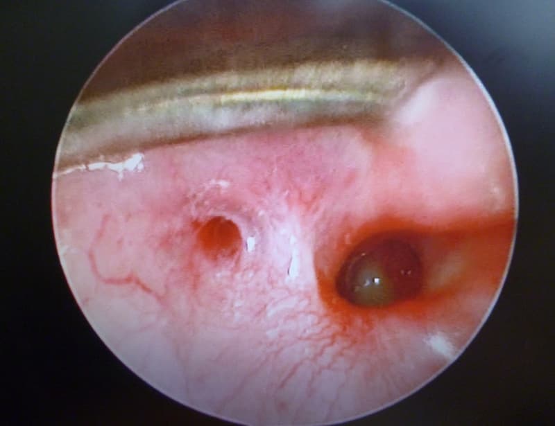
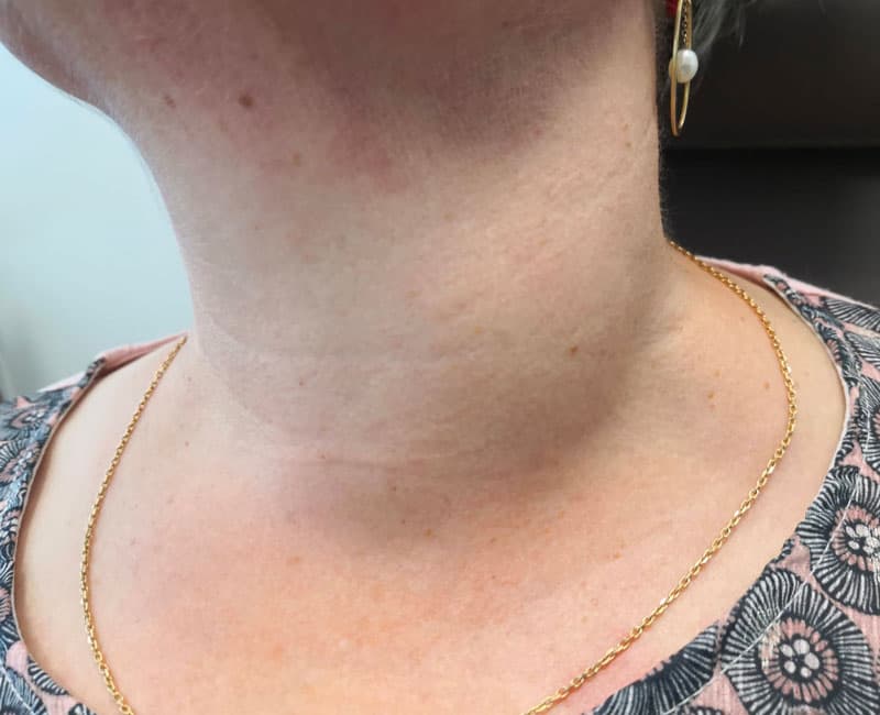
The human body renews itself continuously throughout life, but it is constructed at the embryonic and fetal stages. Organs are created during embryogenesis in pregnancy from primordial tissue layers (endoderm, ectoderm, mesoderm). Cavities and outgrowths formed from these primordial tissue sheets give rise to future organs. These new pre-organic structures result from fusions, sutures and even the disappearance of tissue. Sometimes, anatomical elements that should have disappeared persist in areas of fusion or suture. These persistent features are sometimes not visible at birth, and may even be revealed only in adulthood, but more often than not they are discovered during childhood. They are called “embryonic malformations”. The face and neck are complex anatomical zones where embryonic persistence is not uncommon. They take the form of cysts and fistulas, which can become infected and complicated.
Find out more about surgery for cervico-facial embryonic cysts and fistulas from Dr Delagranda, ENT and cervico-facial surgeon in La Roche sur Yon.
Embryonic malformations: cervico-facial cysts and fistulas
The most common cervico-facial cysts and fistulas are
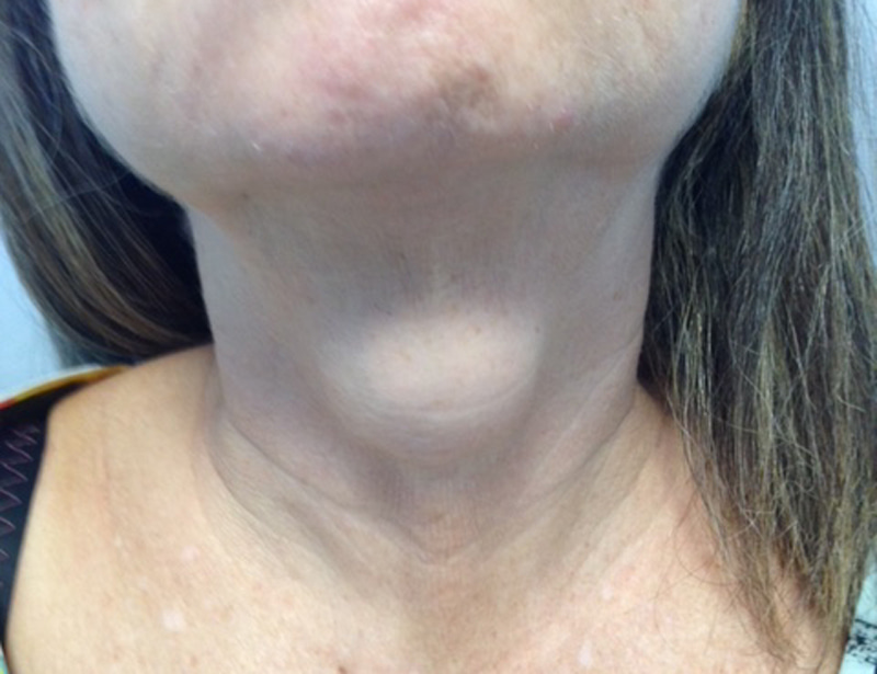
- Thyroglossal duct cysts: a mass located in the middle of the neck, rising towards the chin
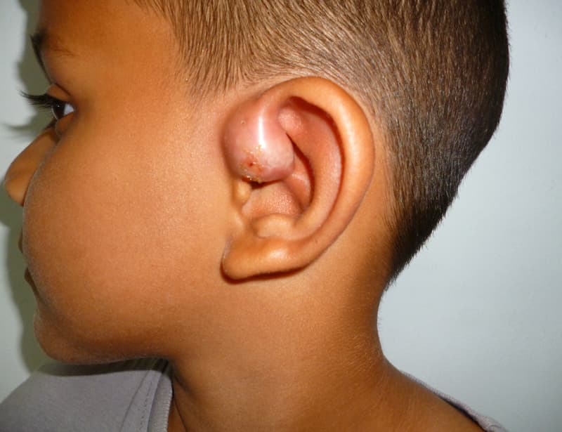
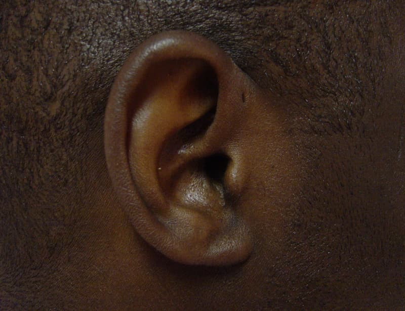
- Pre auricular cysts,pits or fissures: small oozing orifice, with or without a mass, located in front of the ear and above the orifice of the external auditory canal.
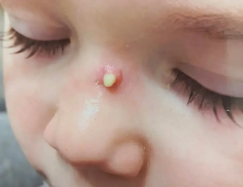
- Cysts and fistulas of the 2nd branchial arch: mass located on the lateral part of the neck, rarely with an orifice above the tonsil.
- 1st branchial arch cysts and fistulas: mass located in the posterior part of the cheek, fistulous orifice in the external auditory canal or cheek.
- Cysts and fistulas of the 3rd and 4th branchial arches: repeated cervical abscesses in the lower neck, simulating suppurative thyroiditis, with fistulous orifice in the hypopharynx at the upper part of the piriform sinus, before the oesophagus, usually on the left side.
- Naso palatine cysts: a mass located in the palate behind the incisors.
- Sternal cysts and fistulas: opening above or at the level of the sternum.
- Cystic lymphangiomas: soft mass of variable size, with one or more cavities, located in all parts of the neck. Treatment is not always surgical: chemical sclerosis is possible in some cases.
Indications, objective and target audience for removal of embryonic cysts and fistulas
Embryonic cysts and fistulas are not always visible at birth, and can even be revealed only in adulthood, but most often they are discovered during childhood.
Removal of embryonic malformations, cysts and fistulas is recommended when they are symptomatic (swelling, oozing, pain, infection), as they do not disappear spontaneously and may even increase in size. Infections are likely to recur many times. The standard treatment is surgical removal.
The aim is to stop repeated infections and deformities in the neck and posterior face.
The different stages of the intervention
The surgical procedure
Only surgery on preauricular cysts in adults can be performed under local anaesthetic on an outpatient basis in 20 minutes. All other cases require general anaesthesia and weekday hospitalization, usually for 2 or 3 days, rarely on an outpatient basis (nasopalatine cyst), as postoperative cervical drainage is required. Operating time varies according to the type of cyst or fistula (minimum 1h – maximum 2h).
Post-surgery recovery period
One day for preauricular cysts, 15-21 days for thyroglossal tract cysts. The patient is provided with a medical leave in connection with the surgery.
Sport is not recommended for the first 21 days, and resumption should be gradual.
Pain is significant in the case of thyroglossal tract cysts. Class II analgesics are recommended.
Post-operative care at home: mouthwash, analgesics, scar care.
Scar: visible horizontal scar in the neck (5 cm).
Complications associated with surgery for embryonic cysts and fistulas
- Tender, painful, inflammatory scar (keloid)
- Hematoma
- Recurrence
Depending on cyst location :
- Loss of sensitivity in earlobe, cheek
- Difficulty swallowing due to pain in the neck or near the ear
- Reduced tongue mobility
- Delayed resumption of eating
- Food misdirection
- Facial paralysis
For further information on indications and risks, please consult the relevant ENT College fact sheets :
➔Exerese fibrochondrome chez l’enfant
➔Exerese kyste tractus thyreoglosse
➔Exerese fistule premiere fente branchiale chez l’enfant
Frequently asked questions
Here is a selection of questions frequently asked by Dr Delagranda’s patients during consultations for embryonic cyst and fistula surgery in La Roche-sur-Yon.
Is there a risk of recurrence?
Yes, these pathologies tend to recur, but careful surgery limits this risk.
Fees and coverage of the operation
Surgery for embryonic cysts and fistulas is covered by the French health insurance system. Contact your mutual insurance company to find out whether any extra fees will be covered.
Do you have a question? Need more information?
Dr Antoine Delagranda will be happy to answer any questions you may have about surgery for embryonic cysts and fistulas. Dr Delagranda is a specialist in ENT surgery at the Clinique Saint Charles in La Roche-sur-Yon in the Vendée.
ENT consultation for embryonic cyst and fistula surgery in Vendée
Dr Antoine Delagranda will be happy to answer any questions you may have about surgery for embryonic cysts and fistulas. Dr Delagranda is a specialist in ENT surgery at the Clinique Saint Charles in La Roche-sur-Yon in the Vendée.


