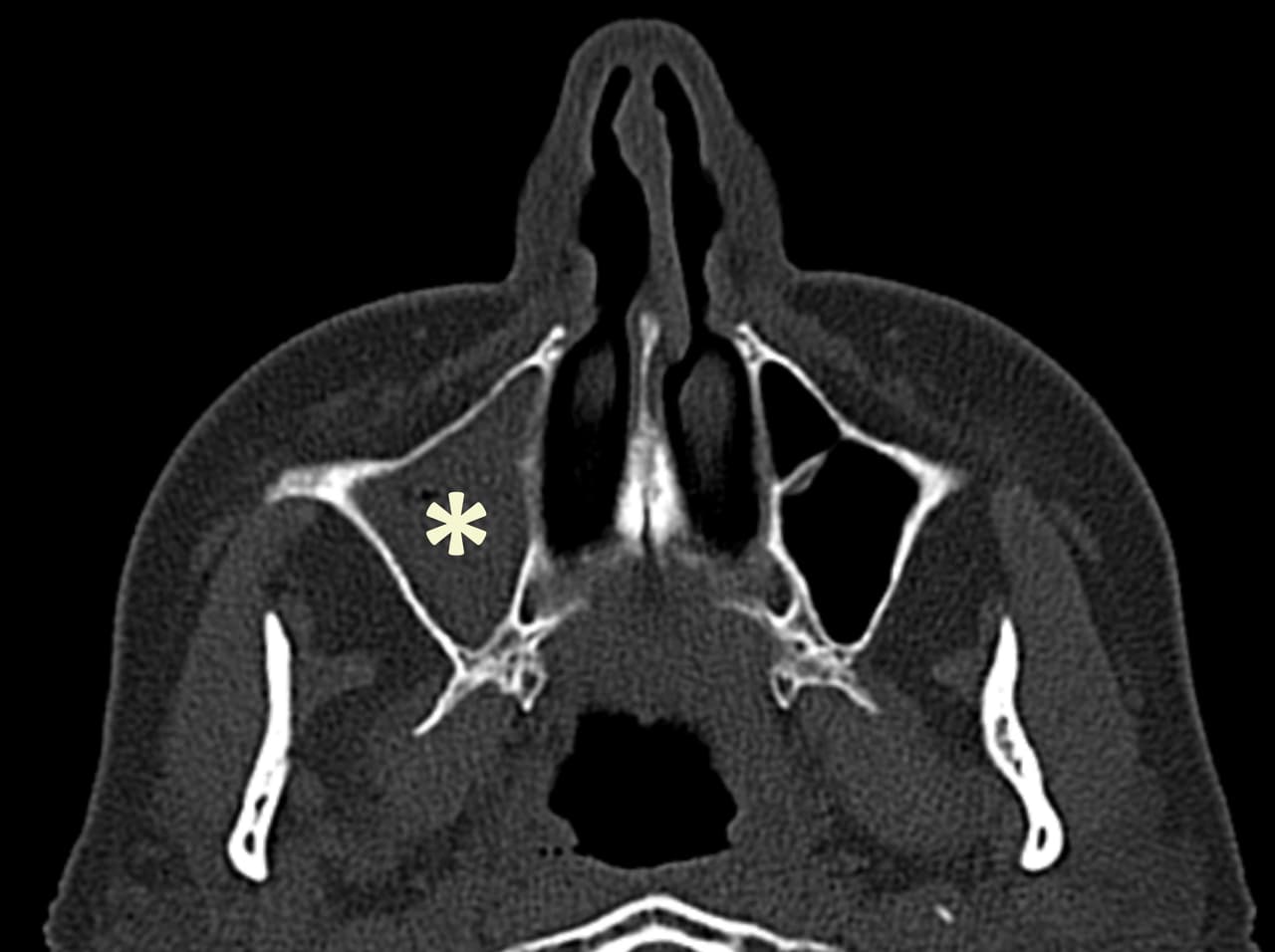
Endonasal meatotomy
Endonasal meatotomy is the opening of the maxillary sinus. It involves enlarging the natural drainage orifice of the sinus, known as the maxillary ostium, to allow the interior of the sinus to be cleaned during the operation, and to ensure good natural cleaning afterwards. Infections and the presence of foreign bodies of dental origin constitute the vast majority of indications for this surgical procedure.
Find out more about endonasal meatotomy below from Dr. Delagranda, ENT and cervico-facial surgeon in La Roche sur Yon.
Maxillary sinus, teeth and fungus
Maxillary sinus
A pneumatized cavity (filled with air from the nasal cavities) in the maxillary bone. Present from birth, unlike other sinuses, it grows until the age of 15, in the shape of a pyramid with a lateral tip. An incomplete septum may be present in the maxillary sinus in 2.4% of cases. It communicates with the nasal cavity via its natural drainage orifice or ostium, located at the level of the intersinonasal septum. In 13.7% of cases, there are accessory ostia known as Giraldès ostia. The maxillary bone alone forms all the walls of the maxillary sinus, except for the intersinonasal septum, which it shares with the inferior turbinate, ethmoidal bone, palatine bone and lacrimal bone.
Sinus teeth
Sinus teeth are the upper premolars and molars, as their roots may be in close contact with the maxillary sinus cavities.
An infectious and inflammatory bacterial lesion at the tips of dental roots, following endocanal infection after caries and pulpitis, forms an apical granuloma that can develop into a radiculodental cyst. The apical granuloma creates osteitis in the vicinity, with disappearance of the bone separating the tooth from the maxillary sinus, leading to development of the infection in the sinus. Dental treatment is mandatory, but sometimes sinusitis fails to heal despite several courses of antibiotics, necessitating a middle meatotomy.
It also happens that a dentist’s treatment of a root canal in one of these sinus teeth results in the passage of dental paste into the sinus floor.
Tooth paste in this open environment may encourage the development of chronic sinus infection, but this is not systematic. The decision to remove this ectopic dental pulp (outside the teeth) and the approach to be taken will depend on the clinical impact, its size and location.
Mycosis
The maxillary sinus is the most frequent site of fungal balls (macroscopically visible fungi), which are responsible for non-invasive, non- or minimally aggressive, extramucosal fungal sinusitis. Aspergillus is the most common type of fungus to be found in fungal slugs, but its detection in culture is not easy (only 30% of cases).
While some fungal slugs develop on dental pulp passed through the sinus, the majority grow from mycotic spores carried by the ambient air and trapped in the sinus. Fungal balls cannot be treated medically, as they are extra-mucosal and voluminous, and must be physically removed by surgery. Diagnosis and treatment depend on a CT scan of the sinuses, which shows characteristic signs (calcifications within an overall opacity of the sinus, thickening of the bony walls).
Exceptionally, but almost always in immunocompromised patients (diabetics, haemopathies, chemotherapy), a fungus (Aspergillus or Mucorales) can penetrate tissues and become invasive. In this case, it is very aggressive, and more extensive surgery accompanied by intravenous antifungal agents (triazole derivatives, amphotericin B) is recommended.
Allergic rhinitis can be triggered by a fungus such as Alternaria, Cladosporium or Aspergillus, and does not require surgery, but medical treatment (antihistamines and inhaled corticoids) and, above all, improvement of the damp living environment. Some works are more at risk: bakers, farmers, market-gardeners, wine-growers, mushroom growers, wood, cheese, sausage and oil workers.
Who is concerned by endonasal meatotomy?
- Adults with fungal sinusitis in the form of solid fungal balls.
- Adults with bacterial sinusitis that is resistant to several antibiotic treatments and despite adequate dental treatment (the cause of maxillary sinusitis is dental in 50% of cases).
- Adults with a resounding foreign body in the maxillary sinus (dental paste, dental implant, shot).
- Adults with a benign tumor in the maxillary sinus.
- Very exceptionally, children with an antrochoanal or Killian polyp.
The case of “polyps” of the sinus fundus, which are rather non-secreting seromucous retention cysts or pseudocysts, should be carefully considered. They are due to chronic inflammation that forms a pseudocyst without a clean wall and lifts the mucosa from the underlying bone wall. Found very frequently (up to 35% of people), they have no pathological character, apart from the exceptional cases where they lead to obstruction. On CT scans, they appear as regular hypodense, homogeneous opacities, convex in shape like a sunset, with no bone lysis or deformation. Often totally isolated, they may also be associated with other pathologies such as dental foci, inflammatory and/or infectious sinus involvement, or anatomical anomalies favoring ostial obstruction. They appear and disappear on successive examinations. Except in exceptional cases of obstruction, they do not constitute an indication for surgery.
When should an endonasal middle meatotomy be performed?
Endonasal meatotomy should be performed in cases of:
- Bacterial sinusitis, despite well-managed medical treatments: blocked nose, running down one side of the nostril and into the throat (nasal obstruction, anterior and posterior rhinorrhea), purulent nose discharge, permanent bad odor (cacosmia), intense pain in the cheekbone, eye or increased heaviness when bending the head forward.
- Fungal bullet sinusitis: with the same signs as bacterial sinusitis, but sometimes completely asymptomatic.
- It is nevertheless recommended to remove this fungus even without symptoms, especially in cases of diabetes, as it constitutes a chronic source of infection.
- Benign tumor of the maxillary sinus.
- Killian’s antrochoanal polyp: progressively blocked nose running down one side of the nostril and into the throat (nasal obstruction, anterior and posterior rhinorrhea) in children as young as 5. The polyp originates in the maxillary sinus, where it is attached, and passes through the intersinuso nasal septum either via the ostium or via an accessory orifice known as the Giraldès orifice, resting in the nasal cavity and choana posteriorly. The bissac-like appearance on CT scan is classic.
Objectives of endonasal middle meatotomy
- Eliminate pain.
- Improve the sensation of a blocked nose.
- Reduce anterior and posterior rhinorrhea and their consequences.
- Eliminate the sensation of bad odor (cacosmia).
- Prevent future complications.
- Remove benign or malignant tumors.

The different stages of the intervention
The surgical procedure
Under general anaesthetic in the operating theatre, the nasal cavities are cleaned with an additional anaesthetic-retractant, the maxillary sinus ostium is located, dangerous anatomical landmarks are palpated to spare them, and a small bone forming part of the ethmoid in the intersinuso-nasal septum – the processus unciforme – is removed. With the unciform process removed, the new, larger opening includes the sinus ostium. This large opening makes it possible to check and clean the inside of the maxillary sinus, using angled optics (30° and 70°) and appropriate forceps and suction. Any fungus, foreign body or tumour is then removed, and the sinus is checked for proper vacuity.
Post-surgery recovery period
In the case of outpatient surgery, the patient usually returns home the same day.
After hospitalization, you’ll need to rest at home for 7 days, and check that there’s no bleeding from the nose or throat.
If necessary, the surgeon will give you 7 to 10 days off work.
Sport is not recommended for the first 15 days, and should be resumed gradually.
Pain is very moderate. It is controlled by Class I analgesics.
Post-operative care at home: nosewash with saline, analgesics, antibiotics if required by your doctor.
Scarring: no visible scar.
Complications associated with endonasal meatotomy
In addition to the risks inherent in any surgery involving general anesthesia, endonasal middle meatotomy presents rare complications:
- Nasal haemorrhage (epistaxis) after the procedure, which is very minor and rapidly subsides with nose-blowing and nose-washing.
- Periorbital hematoma.
- Air trapped in the eyelids (emphysema).
- Infection.
- Tearing.
- Bridles responsible for limiting nasal flow.
Please see the College of ENT’s explanatory sheet on endonasal meatotomy for further explanations and exceptional complications:
Frequently asked questions
Here is a selection of questions frequently asked by Dr Delagranda’s patients during consultations for endonasal meatotomy in La Roche-sur-Yon.
Is surgery compulsory?
Yes, if the indication is well defined, but the surgeon advises and the patient decides.
Is the effect long-lasting?
Yes, there is very little recurrence of mycosis.
Is it painful?
Not really.
Fees and cost of the procedure
Endonasal meatotomy is covered by the French health insurance system. Please contact your health insurance company to find out whether any extra fees will be covered.
Do you have a question? Need more information?
Dr Antoine Delagranda will be happy to answer any questions you may have about endonasal meatotomy. Dr Delagranda is a specialist in ENT surgery at the Clinique Saint Charles in La Roche-sur-Yon in the Vendée.
ENT consultation for endonasal meatotomy in Vendée
Doctor Antoine Delagranda is available to answer any further questions you may have about endonasal middle meatotomy. Dr. Delagranda is a specialist in ENT surgery at the Clinique St-Charles, La Roche sur Yon, France.

