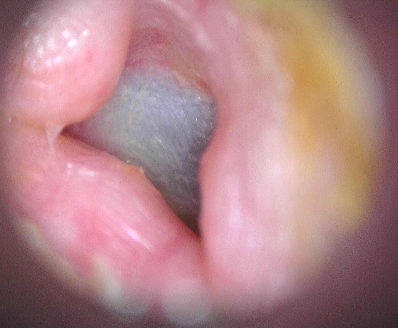
Exostosis or hyperostosis of the external auditory canal
The operation consists in enlarging the diameter of the external auditory canal in the bony part stenosed by abnormal bone formations that have appeared during life.
Find out more about the removal of exostoses of the external auditory canal from Dr Delagranda, ENT and cervico-facial surgeon in La Roche sur Yon.
Who is concerned by the removal of exostosis of the external auditory canal?
Adults with symptoms related to exostoses:
- Adults with repeated otitis externa.
- Adults with an annoying sensation of persistent waterlogging in the ear after water sports or swimming.
- Adults with repeated hearing loss due to earwax or epidermal plugs trapped by exostoses.
- Adults with permanent hearing loss due to totally occlusive exostoses.
- Adults who require middle ear surgery when exostoses prevent access to the eardrum, making surgery directly impossible.
- Adults who have to wear a hearing aid whose earmold is obstructed by exostoses.
Children do not have symptomatic exostoses, as their development is very slow and takes many years. They are therefore not concerned.

External auditory meatus and exostosis
The external auditory canal is located between the pinna on the outside and the eardrum on the inside. It is made up of two parts: a fibrocartilaginous portion making up the outer third (8mm) and a bony portion making up the inner two-thirds (16mm). The bony part is made up of 2 closely linked bones, the temporal bone at the top and back, and the tympanal bone at the bottom and front. In the fibrocartilaginous portion, the skin is thick with sebaceous glands and ceruminous glands that produce earwax, whose role is to protect the skin of the auditory canal thanks to its acid pH. In the bony portion, the skin is very thin and lies directly against the periosteum, which explains why exostosis appear here.
Exostosis or hyperostosis are bone outgrowths that are usually multiple and bilateral, and must be differentiated from an osteoma, which is usually a single benign bone tumor. Exostoses develop very slowly over many years, mainly through repeated exposure to cold water. Studies have shown that they are more common in sportspeople such as surfers, swimmers and divers. Exostosis are found in 0.7% of men and women. Most exostosis are harmless and are discovered incidentally during otoscopic examination of the ear. As exostosis increase in size and obstruct the external auditory canal, they can lead to symptoms.
- A sensation of a full ear, as water can no longer exit the ear properly.
- A blockage of epidermal plugs or earwax, with the sensation of a blocked ear.
- Infection or otitis externa, very painful, due to bacterial or mycological proliferation on macerated skin waste trapped by osteomas.
- Reduced hearing if the obstruction is complete.
Exostoses should only be removed if they cause symptoms.
When should exostosis of the external auditory canal be removed?
Exostosis should be removed in case of:
- Repeated external otitis despite successful medical treatment: severe ear pain (sleepless, throbbing, poorly relieved by paracetamol), increased by pressure on the tragus (pathognomonic sign), sensation of blocked ear.
- Reduced hearing: due to the exostoses themselves, or the cerumen or epidermal plug blocked by the exostoses.
- Annoying sensation of water stagnating in the external auditory canal after every swim.
- Difficulty wearing hearing aids.
- Requires middle ear surgery and removal of exostoses, as they are located on the approach.
The different stages of the intervention
The surgical procedure
There are various reasons for removing exostoses of the external auditory canal:
- Eliminate the pain of otitis externa.
- Eliminate the sensation of stagnant water in the external ear.
- Enable the serene resumption of water sports and scuba diving.
- Improve hearing
- Make it easier to wear an external hearing aid.
- Enable middle ear surgery (myringoplasty, tympanoplasty, otosclerosis).
With general anesthesia, in the operating room, the skin of the auditory canal is peeled off upstream of the exostoses, the bone of the exostoses is exposed and milled until a normal-caliber auditory canal is obtained, protecting the eardrum so as not to damage it. The skin of the canal is placed back over the milled bone, which is applied with a resorbable healing foam held in place by a special hydrophilic canal dressing called POP oto-wick©, soaked in antibiotic liquid.
Post-surgery recovery period
In the case of outpatient hospitalization, the patient is usually discharged home the same day.
After hospitalization, you must rest at home for 7 days.
If necessary, the surgeon will give you 7 to 15 days off work.
Sport is not recommended for the first 15 days, and resumption should be gradual.
Pain is moderate. It is controlled by class I or II analgesics.
Post-operative care at home: daily nursing care of the external scar and antibiotic ear drops several times a day.
Scar: vertical, 5 to 10 mm in front of the ear above the tragus.
Complications associated with the removal of exostosis of the external auditory canal
In addition to the risks inherent in any surgery involving general anaesthesia, the removal of exostoses from the external auditory canal carries the risk of rare or exceptional complications:
- Fibrous scarring, compromising the result.
- Delayed healing.
- Infection.
- Mastication problems.
- Facial paralysis (exceptional).
- Deafness (exceptional).
- Deafness (exceptional paralysis)
Please refer to the College of ENT’s explanatory sheet on external auditory meatus exostosis for further explanations and exceptional complications :
Frequently asked questions
Here is a selection of questions frequently asked by Dr Delagranda’s patients during consultations for the removal of exostoses of the external auditory canal in La Roche-sur-Yon.
Is the operation compulsory?
No, the surgeon advises and the patient decides.
Is the effect lasting?
Yes, any regrowth of exostosis is extremely slow.
Is it painful?
Moderately
Fees and payment for the procedure
Removal of exostoses of the external auditory canal is covered by the French health insurance system. Contact your mutual insurance company to find out whether any extra fees will be covered.
Do you have a question? Need more information?
Doctor Antoine Delagranda will be happy to answer any questions you may have about the removal of exostosis from the external auditory canal. Dr Delagranda is a specialist in ENT surgery at the Clinique Saint Charles, La Roche sur Yon.
ENT consultation for exostosis in Vendée
Doctor Antoine Delagranda will be happy to answer any questions you may have about the removal of exostosis from the external auditory canal. Dr Delagranda is a specialist in ENT surgery at the Clinique Saint Charles, La Roche sur Yon.

