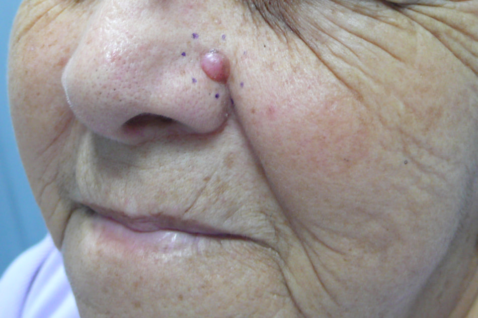
Skin tumor surgery
Cutaneous tumor surgery covers a wide range of surgical procedures, with the common aim of completely removing the diagnosed lesion, with minimal aesthetic and functional after-effects. The modalities of each procedure are tailored to the individual patient, ranging from local anesthesia performed on an outpatient basis to general anesthesia requiring overnight hospitalization – or, more rarely, a few days.
Find out more about skin tumour surgery from Dr Delagranda, ENT and cervico-facial surgeon at La Roche-sur-Yon in the Vendée.
When should skin tumour surgery be considered?
Surgery for skin tumours is mainly performed on adults, more often over the age of 50.
The diagnosis of skin tumors is primarily a clinical one.
Whether the lesion is benign or malignant is generally determined by precise examination of the lesion by a trained physician (usually a dermatologist), but requires histological confirmation (microscopic analysis). Depending on the type of tumour suspected, it is possible to propose either a partial removal, called biopsy, or a complete removal, called excision.
In the case of benign skin tumors, the carcinogenic potential is virtually nil, so the patient’s aesthetic or functional discomfort dictates the surgical indication.
However, if a benign tumour changes in appearance or presents unusual characteristics, it should be considered suspicious, and histological analysis should be carried out to verify the absence of malignant transformation.
In patients presenting lesions suspected of cancer, surgical coverage is mandatory.

The skin
The skin is the heaviest organ in the human body (average 16 pounds/8 kg for an adult). It is a complex organ that envelops the body’s surface, with numerous protective, secretory and sensory functions. It is an organ with remarkable qualities of regeneration, elasticity and plasticity.
It is a stratified organ, i.e. made up of layers, of which there are three main ones:
- the epidermis, the most superficial layer, which acts as a protective barrier against external aggressions (mechanical, ultra-violet, thermal, etc.), but also as a receptor of surrounding information (sensory, immune). These functions are performed by very specific cell populations.
- the dermis, the skin’s intermediate layer, which supports the epidermis. It contains vascular, lymphatic and nervous networks, as well as sweat glands and hair follicles. Made up of connective tissue, this layer is responsible for the skin’s elasticity, and with aging, for the appearance of wrinkles.
- the hypodermis, the deepest, “subcutaneous” layer, which connects the skin to the underlying organs via loose connective tissue, allowing it to slide in relation to the deep plane. It contains adipose cells or adipocytes, in greater or lesser quantities depending on location and individual.
Skin lesions and tumours
The skin is a complex organ made up of a wide variety of cell types, which explains the multiplicity of skin lesions.
The term tumour refers to the excessive multiplication of cells, either normal (benign tumour) or abnormal (malignant tumour), resulting in a lump or swelling of varying size.
Benign tumours develop locally. The main benign skin tumours are nevi or moles developed from melanocytes, keratotic lesions (seborrhoeic or actinic) developed from keratinocytes, sebaceous cysts developed from sebaceous glands and lipomas which develop in the fat cells of the hypodermis.
Malignant tumours, commonly known as “cancer”, tend to invade neighbouring tissues and migrate to other parts of the body (metastases). Malignant skin tumours are the most common malignant tumours in adults. The incidence of these tumours is rising, with more than 65,000 patients in France in 2010 suffering from skin tumours in the cephalic region (head and neck). This frequency is explained by the ageing of the population and the close link with sun exposure in the development of skin tumours.
Main malignant skin tumours
The main malignant cutaneous tumors also include a wide variety of histological lesions, of which we will only detail here:
- Cutaneous carcinomas or epitheliomas develop from epidermal cells called keratinocytes. These cells, stacked one on top of the other in several layers, are responsible for the skin’s physical barrier function. They are particularly sensitive to exposure to chronic or intermittent solar UV radiation (“sunburn”), which causes local genetic abnormalities leading to transformation into cancer.
A distinction is made between- basal cell carcinomas (BCCs), the most common, are slow-growing tumors that are essentially local, never metastasizing, but can be destructive. The safety margin for cutaneous excision is 5 mm to avoid recurrence. It is preferable to book these tumors early to avoid major mutilation.
- squamous cell carcinoma (SCC) or squamous cell carcinoma (SCC), which are more aggressive, can metastasize and often arise on a precancerous lesion. An injected cervical CT scan is recommended to look for suspicious lymph nodes, which may need to be removed as part of a lymph node curage. The safety margin for skin resection is between 6 and 10 mm.
- Melanomas are skin tumors that develop from epidermal cells called melanocytes. Melanocytes protect the body from the sun’s UV rays by producing pigments (eumelanin and pheomelanin), which they distribute to all the keratinocytes that make up the epidermis. The quantity of these pigments, which genetically determine the color of our skin, varies slightly with exposure to the sun. The aggressiveness of melanomas depends on their thickness: the Breslow index. The safety margin will be adapted to the Breslow result. In order to determine whether lymph nodes have been invaded, beyond 0.8mm of Breslow, the sentinel lymph node technique is commonly used. Extension workup with brain MRI and PET scan is recommended. The therapeutic decision is taken in a multidisciplinary consultation involving dermatologists, oncologists, radiotherapists and surgeons. In addition to surgery, chemotherapy or immunotherapy may be proposed in the case of metastases. Melanomas are skin tumors that affect all ages, although they are exceptional in children before puberty. In France, the incidence is estimated at 5-10 new cases/100,000 inhabitants/year. It most often occurs on fair skins, and intermittent, intense exposure (“sunburn”) is a major risk factor. A family history is found in 10% of cases. It’s important to point out that most melanomas arise without precursors (“de novo”), but any recent change in a mole or nevus should prompt a specialist medical consultation.
Objectives and principles of the intervention
The aim of surgical coverage for both benign and malignant skin tumors is to remove the entire tumor, with a margin of healthy skin, in order to carry out a complete histological analysis and repair the loss of substance.
In certain cases, depending on the size or location of the lesion, the surgeon may propose a biopsy (partial sampling), to identify the type of tumor and guide the surgical procedure.
The basic principle of surgical intervention for skin lesions of the face and neck is spindle-shaped excision followed by direct suturing: the surgeon makes an elliptical incision around the lesion, leaving a gap of healthy skin or margins, then sutures the 2 incision edges together. Although the resulting scar is often longer than the initial tumour, it is oriented parallel to the tension lines of the skin on the face and neck, ensuring the best possible aesthetic result.
When a spindle-shaped excision with direct closure is not feasible, due to the location or size of the lesion, the surgeon has a wide range of options at his disposal: After excision of the tumor, he can perform :
- a skin graft taken from another, less visible part of the body. The harvested area will generally require simple dressing by a nurse.
- a local plasty skin flap: by making precise incisions in the excess healthy skin in the vicinity of the lesion, it is possible, at the cost of a slightly larger scar, to repair a large loss of substance with very acceptable aesthetic results. Nursing dressings at home once or twice a day are necessary for the first two weeks.
- If the loss of substance is too great or too deep, the surgeon may need to perform a “free flap” repair: a segment of skin is harvested from another area of the body, at varying depths, sometimes right down to the muscle, along with the vessels responsible for its vascularization, to reconstruct the loss of substance on the face or neck. There are a large number of preferred harvesting areas (forearm, thigh, lateral part of the back), which can be adapted according to the reconstruction required and the surgeon’s experience. The harvested areas are either closed by direct suture or skin grafting. For this type of operation, a general anaesthetic and several nights’ hospitalization are required to ensure smooth progress and reliable results. Your surgeon can help you in this process.
In certain special cases of suspected tumors with poorly defined limits, or recurrent malignant tumors, repair of the loss of substance is carried out after complete analysis of the excision of the lesion, in a second stage of surgery.
Complications associated with skin tumour surgery
As with any surgical procedure, there may be some discomfort, notably a feeling of tension in the operated area, but the post-operative period is generally painless.
Early signs:
Bleeding: a more or less clear flow of blood through the scar, usually of small volume, controlled by dressing.
Post-operative hematoma: an organized blood clot inside the scar, usually small in size. In most cases, it requires only simple monitoring. Occasionally, it may require a new operation for evacuation.
Infection: Inherent to any surgical procedure, infection is evidenced by an opening in the scar and/or pain in the area and/or a dirty, purulent discharge and/or reddening of the skin. It may be encouraged by poor suture tolerance or poor local care. Antibiotics are often prescribed, and scarring results may be altered.
Faulty or delayed healing: This type of complication is more frequent in smokers and poorly-balanced diabetics. Loose stitches may also occur. As a rule, treatment is based on careful dressing. Occasionally, re-intervention may be required, with a new skin graft or flap.
Nerve injuries: Stretch-injury or even sectioning of sensory or motor nerves is rare in face and neck surgery, particularly in the case of sectioning of one or more branches of the facial nerve. Sometimes, the carcinological objective of total removal of the malignant tumor requires sectioning of a motor branch, resulting in generally limited facial asymmetry.
Delayed signs:
Scarring anomaly: the healing process lasts several months, with scars evolving up to 1 or 2 years after surgery. Skin scars may become or remain inflammatory for several months, possibly warranting local corticosteroid injections. Very large, progressive hypertrophic scars (keloids) are very rare on the face, except behind the ears, and may justify complementary treatments such as pressotherapy.
Re-operation and re-operation: in the case of malignant skin tumours, in the event of incomplete removal or insufficient healthy skin margins, re-operation is necessary to restore the margin. Moreover, the main risk for a patient who has had a malignant tumor is that of local or locoregional recurrence (lymph node area). Prolonged surveillance is usually carried out by a dermatologist.
Frequently asked questions
Here is a selection of questions frequently asked by Dr Delagranda’s patients during consultations for skin tumours in La Roche-sur-Yon.
Is a scar compulsory after the removal of a skin tumor, even a benign one?
Surgical removal requires a full-thickness incision in the skin, generating a de facto scar, which cannot disappear completely, regardless of the surgeon’s skill.
I'm taking anticoagulants or platelet anti-aggregants (such as Aspégic©, Kardégic©, Plavix©, Duoplavin©, Préviscan©, ...), do I have to stop them for the operation?
Except in special cases, you should stop taking your medication a few days before and after the operation to limit the risk of bleeding and hematoma. Your surgeon or anaesthetist will inform you of the time limits to be respected, depending on your treatment and the procedure to be performed, during his or her preoperative consultation.
How long do I need to protect my scar?
Once the sutures have been removed and your surgeon has checked that the scar has healed properly, you will need systematic sun protection using sunscreen for a period of 1 year. Healing creams can also be recommended by your surgeon or pharmacy, to be applied daily for the first few months. This care is designed to ensure the best possible aesthetic result for your scar, which can be modified up to 1 or even 2 years after surgery.
I had skin resection surgery. Will I need further treatment?
In the case of malignant skin tumors, radiotherapy and/or chemotherapy are sometimes combined. Histological analysis will guide this additional coverage.
Are neck lymph nodes systematically removed?
No. Only for certain types of malignant tumors, for advanced tumors or when there is doubt about lymph node involvement on clinical examination, generally after an imaging examination (cervical CT and/or PET-Scan and/or cervical MRI).
Fees and surgical coverage
Surgery for skin tumors is covered by the French health insurance system. Contact your mutual insurance company to find out how much coverage there is for any extra fees.
Do you have a question? Need more information?
Dr Antoine Delagranda will be happy to answer any questions you may have about skin tumour surgery. Dr Delagranda is a specialist in ENT surgery at the Clinique Saint Charles in La Roche-sur-Yon in the Vendée.
ENT consultation for skin tumours in Vendée
Doctor Antoine Delagranda is available to answer any further questions you may have about skin tumor surgery. Dr. Delagranda is a specialist in ENT surgery at the Clinique St-Charles in La Roche sur Yon, France.

